临床荟萃 ›› 2024, Vol. 39 ›› Issue (6): 531-536.doi: 10.3969/j.issn.1004-583X.2024.06.008

• 论著 • 上一篇    下一篇

对比机械通气重症肺炎及普通肺炎患者右心功能及右心室-肺动脉耦联的斑点追踪超声评价

燕亚茹1,2, 赵浩天1,3a, 张捷思1,3a, 王晓娜3a, 赵鹤龄3b()   

  1. 1.河北医科大学研究生学院,河北 石家庄 050017
    2.石家庄市人民医院 超声科,河北 石家庄 050051
    3.河北省人民医院 a.超声科;b.重症医学科,河北 石家庄 050051
  • 收稿日期:2023-11-24 出版日期:2024-06-20 发布日期:2024-07-18
  • 通讯作者: 赵鹤龄,Email:epson7777777@163.com
  • 基金资助:
    河北省医学科学研究课题计划项目——心肺超声对重症右心衰竭患者心室动脉耦联效应的研究(20231609);河北省医学科学研究课题计划项目——急重症超声联合心肺交互作用对急重症疾病的诊断价值(20211264)

Ultrasound evaluation of speckle tracking imaging on right ventricular function and right ventricular pulmonary artery coupling in severe pneumonia underwent mechanical ventilation versus common pneumonia

Yan Yaru1,2, Zhao Haotian1,3a, Zhang Jiesi1,3a, Wang Xiaona3a, Zhao Heling3b()   

  1. 1. Graduate School of Hebei Medical University, Shijiazhuang 050017, China
    2. Department of Ultrasound, Shijiazhuang People's Hospital, Shijiazhuang 050051, China
    3a. Department of Ultrasound; b. Department of Intensive Care Unit, Hebei General Hospital, Shijiazhuang 050051, China
  • Received:2023-11-24 Online:2024-06-20 Published:2024-07-18
  • Contact: Zhao Heling, Email: epson7777777@163.com

摘要:

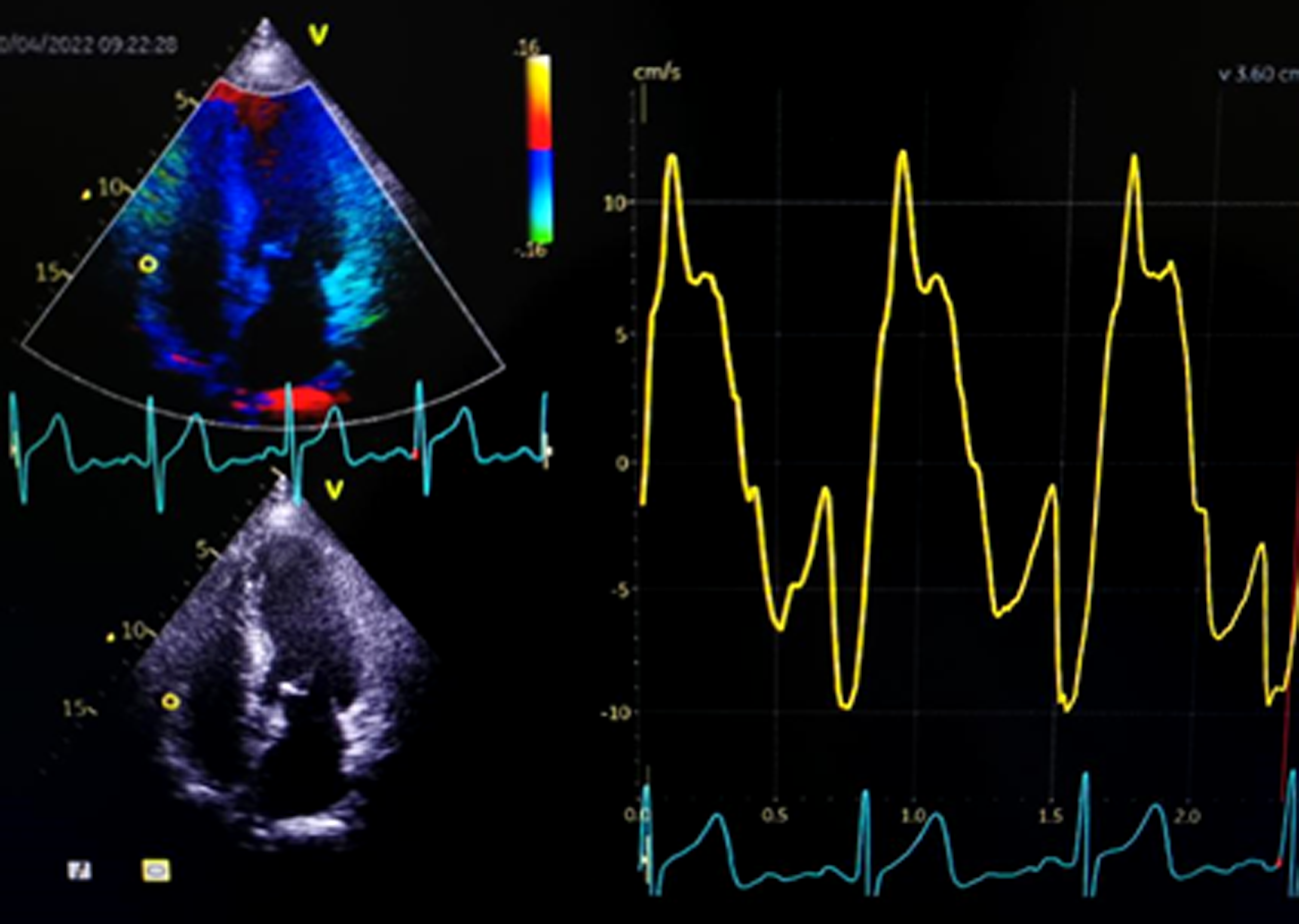
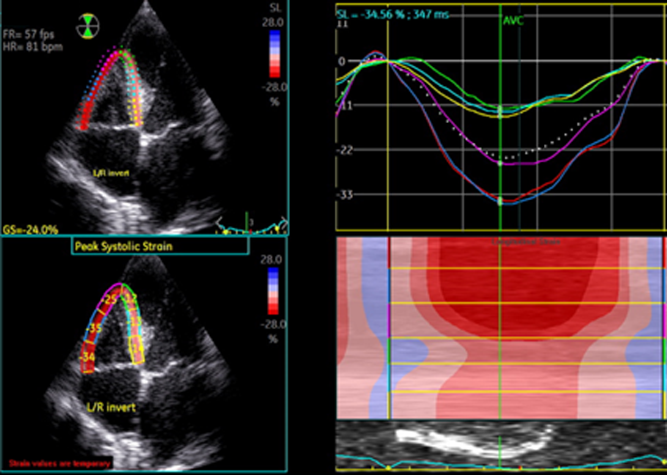
目的 对比机械通气重症肺炎及普通肺炎患者的右心功能超声指标及右心室-肺动脉耦联指标的二维斑点追踪成像(2D-STI)评估价值。方法 选取66例肺炎患者,根据重症肺炎临床标准分为重症肺炎组25例和普通肺炎组41例,另纳入20例健康人为对照组进行超声心动图检查。于心尖四腔心切面获取右心室中段内径(RVD)、三尖瓣环收缩期位移(TAPSE)、三尖瓣环收缩期峰速度(S’)、三尖瓣反流峰值流速(TRV);经剑突下获取下腔静脉(IVC)长轴内径(IVCD)并计算呼吸变异率(IVCV)并估测右房压(RAP),并计算肺动脉收缩压(PASP)。右心室-肺动脉耦联指标以三尖瓣环收缩期位移(tricuspid annular plane systolic excursion,TAPSE)和肺动脉收缩压(pulmonary artery systolic pressure,PASP)的比值(TAPSE/PASP)表示。应用二维斑点追踪成像(two-dimensional speckle tracking imaging,2D-STI)技术获取右心室游离壁整体应变(RVLSfw),基底段应变(RVLSbas)、中段应变(RVLSmid)、心尖段应变(RVLSapi)。将RVLSfw纳入右心室-肺动脉耦联中获取新指标RVLSfw/PASP,比较组间差异并做相关性分析。结果 重症肺炎组RVD、IVCD、TRV、PASP均高于普通肺炎组和对照组,IVCV、TAPSE均低于普通肺炎组和对照组(P<0.05),重症肺炎组S’低于普通肺炎组(P<0.05);普通肺炎组RVD、IVCD高于对照组(P<0.05)。2D-STI指标:重症肺炎组RVLSfw、RVLSbas和RVLSapi均低于普通肺炎组和对照组(P<0.05),普通肺炎组和对照组之间该指标无统计学意义(P>0.05);右心室-肺动脉耦联指标:重症肺炎组TAPSE/PASP、S’/PASP和RVLSfw/PASP均低于普通肺炎组和对照组,普通肺炎组和对照组之间该指标差异无统计学意义(P>0.05)。相关性分析显示:RVLSfw/PASP和TAPSE/PASP呈强相关(r=0.927,P<0.05)。结论 2D-STI指标和右心室-肺动脉耦联均对接受机械通气的重症肺炎患者的右心功能评估有一定价值,RVLSfw/PASP可作为评估右心室-肺动脉耦联的可靠指标。

关键词: 重症肺炎, 心室功能,右, 二维斑点追踪成像, 右心室-肺动脉耦联, 三尖瓣环收缩期位移

Abstract:

Objective To analyze ultrasound evaluation of two-dimensional speckle tracking imaging (2D-STI) on right ventricular (RV) function and RV- pulmonary artery coupling in severe pneumonia underwent mechanical ventilation versus common pneumonia. Methods Sixty-six patients of pneumonia were divided into severe pneumonia group (group S, 25 cases) and general pneumonia group (group G, 41 cases), and 20 healthy people were enrolled as control group. Ultrasonic parameters were obtained in apical four-chamber images, including RV diameter (RVD), tricuspid annular plane systolic excursion (TAPSE), tricuspid lateral annular peak systolic velocity (S'), tricuspid regurgitation velocity (TRV). Inferior vena cava diameter (IVCD) and inferior vena cava variability (IVCV), and the RV pressure (RVP) were estimated, and pulmonary artery systolic pressure (PASP) was calculated. The RV-pulmonary artery coupling index was represented by TAPSE/PASP. The 2D-STE was performed to observe strain parameters, including right ventricular longitudinal strain in the free wall (RVLSfw), right ventricular longitudinal strain in the basal segment (RVLSbas), right ventricular longitudinal strain in the middle segment (RVLSmid), and right ventricular longitudinal strain in the apical segment (RVLSapi). The new index RVLSfw/PASP was obtained by incorporating RVLSfw into the RV-pulmonary artery coupling. The between groups differences and the correlation were assessed. Results RVD, IVCD, TRV and PASP in group S were significantly higher than those in group G and control group, while IVCV and TAPSE were significantly lower (P<0.05). S’ in group S was significantly lower than that in group G (P<0.05). RVD and IVCD in group G were significantly higher than those in control group (P<0.05). 2D-STI indexes including RVLSfw, RVLSbas and RVLSapi in group S were significantly lower than those in group G and control group (P<0.05). There was no statistically significant difference in 2D-STI indexes between group G and control group (P>0.05). RV-pulmonary artery coupling indexes including TAPSE/PASP, S'/PASP and RVLSfw/PASP in group S were significantly lower than those in group G and control group, and there was no statistical significance in RV-pulmonary artery coupling indexes between group G and control group (P>0.05). RVLSfw/PASP was strongly correlated with TAPSE/PASP (r=0.927, P<0.05). Conclusion Both 2D-STI index and RV-pulmonary artery coupling have certain value in evaluating the RV function of patients with severe pneumonia underwent mechanical ventilation. RVLSfw/PASP can be used as a reliable index to evaluate RV-pulmonary artery coupling.

Key words: severe pneumonia, ventricular function,right, two-dimensional speckle tracking imaging, right ventricle-pulmonary artery coupling, tricuspid annular plane systolic excursion

中图分类号: