Clinical Focus ›› 2022, Vol. 37 ›› Issue (11): 1012-1016.doi: 10.3969/j.issn.1004-583X.2022.11.009
Previous Articles Next Articles
Diagnostic value of monosaccharide in peritoneal dialysis-associated peritonitis
Gong Zhiqing, Chen Yipeng, Lu Dongmei, Xin Lili, Guo Jing, Xing Guangqun( )
)
- Department of Nephrology,the Affiliated Hospital of Qingdao University,Qingdao 266555,China
-
Received:2022-07-29Online:2022-11-20Published:2023-01-02 -
Contact:Xing Guangqun E-mail:xinggq@qdu.edu.cn
CLC Number:
Cite this article
Gong Zhiqing, Chen Yipeng, Lu Dongmei, Xin Lili, Guo Jing, Xing Guangqun. Diagnostic value of monosaccharide in peritoneal dialysis-associated peritonitis[J]. Clinical Focus, 2022, 37(11): 1012-1016.
share this article
Add to citation manager EndNote|Ris|BibTeX
URL: https://huicui.hebmu.edu.cn/EN/10.3969/j.issn.1004-583X.2022.11.009
| 项目 | 无腹膜炎组 ( | 革兰阳性菌腹膜炎组 ( | 革兰阴性菌腹膜炎组 ( | χ2值 | |
|---|---|---|---|---|---|
| 男[例(%)] | 27(51.92) | 9(37.50) | 13(92.86) | 11.240 | 0.004 |
| 年龄(岁) | 44.90±15.58 | 50.96±16.91 | 64.71±6.88 | 9.759 | <0.01 |
| BMI(kg/m2) | 25.54±4.40 | 22.92±2.73* | 22.96±5.42* | 4.227 | 0.018 |
| 总蛋白(g/L) | 57.60±8.40 | 52.25±8.40 | 55.60±7.91 | 3.333 | 0.04 |
| 白蛋白(g/L) | 32.57±5.91 | 26.39±5.58* | 24.24±6.92* | 15.476 | <0.01 |
| 尿素(mmol/L) | 23.45±8.73 | 18.07±4.00 | 16.29±4.86 | 7.891 | 0.01 |
| 肌酐(μmol/L) | 875.14±364.85 | 606.70±336.05 | 439.17±114.01 | 12.025 | <0.01 |
| 尿酸(μmol/L) | 388.66±131.35 | 302.85±66.01 | 367.23±164.61 | 3.944 | 0.023 |
| 血钾(mmol/L) | 4.16±0.68 | 3.83±0.45 | 4.50±0.60 | 5.439 | 0.006 |
| 血钠(mmol/L) | 141.20±2.47 | 139.24±3.16* | 136.33±2.36* | 19.691 | <0.01 |
| 血钙(mmol/L) | 2.07±0.20 | 1.97±0.16* | 1.89±0.21* | 5.837 | 0.004 |
| 血磷(mmol/L) | 1.96±0.64 | 1.63±0.47* | 1.09±0.35* | 13.673 | <0.01 |
| 血糖空腹(mmol/L) | 4.73±1.02 | 5.36±1.38 | 9.95±3.36 | 51.203 | <0.01 |
| 血红蛋白(g/L) | 98.10±19.58 | 97.21±11.13 | 106.64±18.15 | 1.523 | 0.224 |
| 淋巴细胞计数(109/L) | 1.41±0.62 | 1.10±0.52 | 1.08±0.50 | 3.361 | 0.039 |
| 血小板计数(109/L) | 181.00±77.52 | 228.46±91.64 | 221.43±106.19 | 3.005 | 0.055 |
| CRP(mg/L) | 1.03(0.50,8.17) | 39.63(18.81,61.22)* | 61.85(36.78,93.60)* | 47.045 | <0.01 |
| PTH(pg/ml) | 356.00±394.15 | 194.47±163.49 | 188.92±107.73 | 2.543 | 0.085 |
| 甘油三酯(mmol/L) | 1.86±1.25 | 1.97±1.62 | 1.21±0.69 | 1.247 | 0.293 |
| 总胆固醇(mmol/L) | 4.88±1.71 | 4.90±1.40 | 4.07±2.73 | 0.791 | 0.457 |
| 组别 | 例数 | 甘露糖 | 葡萄糖 | 岩藻糖 |
|---|---|---|---|---|
| 无腹膜炎组 | 52 | 0.50(0.32,0.73) | 9.30(5.74,15.64) | 5.19(3.59,8.53) |
| 革兰阳性菌腹膜炎组 | 24 | 0.24(0.18, 0.30)*# | 5.02(2.69, 8.52)* | 2.01(1.07, 4.04)* |
| 革兰阴性菌腹膜炎组 | 14 | 0.46(0.33, 0.74) | 3.24(1.98, 6.31)* | 2.48(0.79, 3.17)* |
| 24.010 | 19.875 | 26.918 | ||
| <0.01 | <0.01 | <0.01 |
| 病原菌种类 | 例数 | 构成比(%) |
|---|---|---|
| 革兰阳性菌 | 24 | 63.16 |
| 耳葡萄球菌 | 1 | 2.63 |
| 表皮葡萄球菌 | 5 | 13.16 |
| 金黄色葡萄球菌 | 2 | 5.26 |
| 人葡萄球菌 | 3 | 7.89 |
| 苏云金芽孢杆菌 | 3 | 7.89 |
| 头状葡萄球菌 | 1 | 2.63 |
| 解没食子酸链球菌解没食子酸亚种 | 2 | 5.26 |
| 鹑鸡肠球菌 | 3 | 7.89 |
| 戈登链球菌 | 2 | 5.26 |
| 唾液链球菌 | 1 | 2.63 |
| 革兰阳性菌 | 1 | 2.63 |
| 革兰阴性菌 | 14 | 36.84 |
| 大肠埃希菌 | 3 | 7.89 |
| 革兰阴性菌 | 6 | 15.79 |
| 肺炎克雷伯菌肺炎亚种 | 2 | 5.26 |
| 铜绿假单胞菌 | 3 | 7.89 |
| 因素 | 回归 系数 | 标准误 | Wald χ2值 | 95% | |||
|---|---|---|---|---|---|---|---|
| 下限 | 上限 | ||||||
| 葡萄糖浓度 | -0.046 | 0.069 | 0.435 | 0.509 | 0.955 | 0.834 | 1.094 |
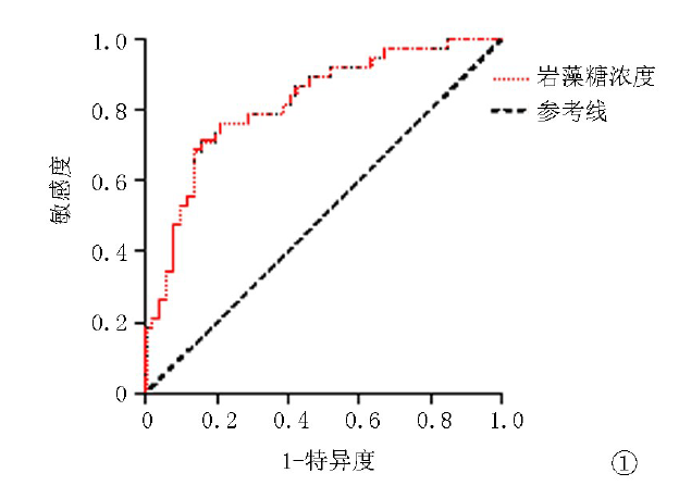
| 岩藻糖浓度 | -0.405 | 0.155 | 6.822 | 0.009 | 0.667 | 0.492 | 0.904 |
| 因素 | 回归 系数 | 标准误 | Wald χ2值 | 95% | |||
|---|---|---|---|---|---|---|---|
| 下限 | 上限 | ||||||
| 葡萄糖浓度 | -0.046 | 0.069 | 0.435 | 0.509 | 0.955 | 0.834 | 1.094 |
| 岩藻糖浓度 | -0.405 | 0.155 | 6.822 | 0.009 | 0.667 | 0.492 | 0.904 |
| 因素 | 赋值说明 |
|---|---|
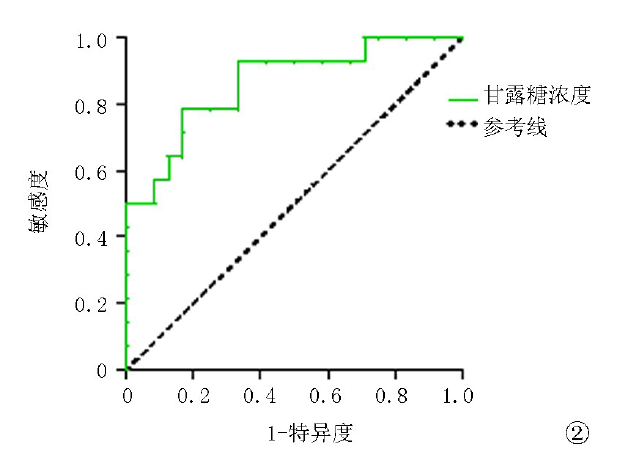
| 甘露糖浓度 | 0=≤0.345,1=>0.345 |
| 因素 | 赋值说明 |
|---|---|
| 甘露糖浓度 | 0=≤0.345,1=>0.345 |
| 因素 | 回归 系数 | 标准误 | Wald χ2值 | 95% | |||
|---|---|---|---|---|---|---|---|
| 下限 | 上限 | ||||||
| 甘露糖浓度 | -2.187 | 0.791 | 7.639 | 0.006 | 0.112 | 0.024 | 0.529 |
| 因素 | 回归 系数 | 标准误 | Wald χ2值 | 95% | |||
|---|---|---|---|---|---|---|---|
| 下限 | 上限 | ||||||
| 甘露糖浓度 | -2.187 | 0.791 | 7.639 | 0.006 | 0.112 | 0.024 | 0.529 |
| [1] |
Li PK, Chow KM, Cho Y, et al. ISPD peritonitis guideline recommendations: 2022 update on prevention and treatment[J]. Perit Dial Int, 2022, 42(2):110-153.
doi: 10.1177/08968608221080586 URL |
| [2] | 杨琼琼, 余学清. 腹膜透析相关感染的防治指南[J]. 中华肾脏病杂志, 2018, 34(2):139-148. |
| [3] |
Yan H, Ma D, Yang S, et al. Effluent lipopolysaccharide is a prompt marker of peritoneal dialysis-related gram-negative peritonitis[J]. Perit Dial Int, 2020, 40(5):455-461.
doi: 10.1177/0896860819896134 URL |
| [4] |
Campanero-Rhodes MA, Palma AS, Menendez M, et al. Microarray strategies for exploring bacterial surface glycans and their interactions with glycan-binding proteins[J]. Front Microbiol, 2019, 10:2909.
doi: 10.3389/fmicb.2019.02909 pmid: 32010066 |
| [5] |
Milan Manani S, Virzì GM, Giuliani A, et al. Lipopolysaccharide evaluation in peritoneal dialysis patients with peritonitis[J]. Blood purification, 2020, 49(4):434-439.
doi: 10.1159/000505388 pmid: 31914448 |
| [6] |
Worasilchai N, Leelahavanichkul A, Kanjanabuch T, et al. (1-->3)-beta-D-glucan and galactomannan testing for the diagnosis of fungal peritonitis in peritoneal dialysis patients, a pilot study[J]. Med Mycol, 2015, 53(4):338-346.
doi: 10.1093/mmy/myv007 pmid: 25851260 |
| [7] |
Desmarais SM, De Pedro MA, Cava F, et al. Peptidoglycan at its peaks: how chromatographic analyses can reveal bacterial cell wall structure and assembly[J]. Mol Microbiol, 2013, 89(1):1-13.
doi: 10.1111/mmi.12266 pmid: 23679048 |
| [8] |
Trezzi JP, Galozzi S, Jaeger C, et al. Distinct metabolomic signature in cerebrospinal fluid in early parkinson's disease[J]. Mov Disord, 2017, 32(10):1401-1408.
doi: 10.1002/mds.27132 URL |
| [9] |
Zhang Y, Ma X, Zhang L. Glycosaminoglycan Quality Control by Monosaccharide Analysis[J]. Methods Mol Biol, 2022, 2303:297-306.
doi: 10.1007/978-1-0716-1398-6_24 pmid: 34626388 |
| [10] |
Rudolph WW, Gunzer F, Trauth M, et al. Comparison of VITEK 2, MALDI-TOF MS, 16S rRNA gene sequencing, and whole-genome sequencing for identification of Roseomonas mucosa[J]. Microb Pathog, 2019, 134:103576.
doi: 10.1016/j.micpath.2019.103576 URL |
| [11] |
Prasad N, Singh K, Gupta A, et al. Isolation of bacterial DNA followed by sequencing and differing cytokine response in peritoneal dialysis effluent help in identifying bacteria in culture negative peritonitis[J]. Nephrology (Carlton), 2018, 23(2):148-154.
doi: 10.1111/nep.12969 URL |
| [12] |
van Hougenhouck-Tulleken WG, Lebre PH, Said M, et al. Bacterial pathogens in peritoneal dialysis peritonitis: Insights from next-generation sequencing[J]. Perit Dial Int., 2020, 40(6):581-586.
doi: 10.1177/0896860820908473 URL |
| [13] |
Alafeef M, Moitra P, Pan D. Nano-enabled sensing approaches for pathogenic bacterial detection[J]. Biosens Bioelectron, 2020, 165:112276.
doi: 10.1016/j.bios.2020.112276 URL |
| [14] | 吴惠娟, 韦曲星, 张盛昔, 等. 甘露糖对巨噬细胞炎症反应的双向调节作用[J]. 中山大学学报(医学科学版), 2020, 41(4):549-557. |
| [15] | Xu XL, Zhang P, Shen YH, et al. Mannose prevents acute lung injury through mannose receptor pathway and contributes to regulate PPARgamma and TGF-beta1 level[J]. Int J Clin Exp Pathol, 2015, 8(6):6214-6224. |
| Viewed | ||||||
|
Full text |
|
|||||
|
Abstract |
|
|||||