临床荟萃 ›› 2022, Vol. 37 ›› Issue (12): 1122-1126.doi: 10.3969/j.issn.1004-583X.2022.12.011

• 论著 • 上一篇    下一篇

TFEB易位肾细胞癌1例并文献复习

何晨冬1, 葛凤林2a, 杨巍2b()   

  1. 1.蚌埠医学院第一附属医院 放射科,安徽 蚌埠 233004
    2.南京中医药大学附属医院 江苏省中医院 a.病理科;b.放射科,江苏 南京 210029
  • 收稿日期:2022-04-14 出版日期:2022-12-20 发布日期:2023-01-18
  • 通讯作者: 杨巍 E-mail:youngwei0713@163.com

TFEB translocation renal cell carcinoma: A case and literature review

He Chendong1, Ge Fenglin2a, Yang Wei2b()   

  1. 1. Department of Radiology,the First Affiliated Hospital of Bengbu Medical College,Bengbu 233004,China
    2a. Department of Pathology; 2b.Department of Radiology,Affiliated Hospital of Nanjing University of Chinese Medicine,Jiangsu Province Hospital of Chinese Medicine,Nanjing 210029,China
  • Received:2022-04-14 Online:2022-12-20 Published:2023-01-18
  • Contact: Yang Wei E-mail:youngwei0713@163.com

摘要:

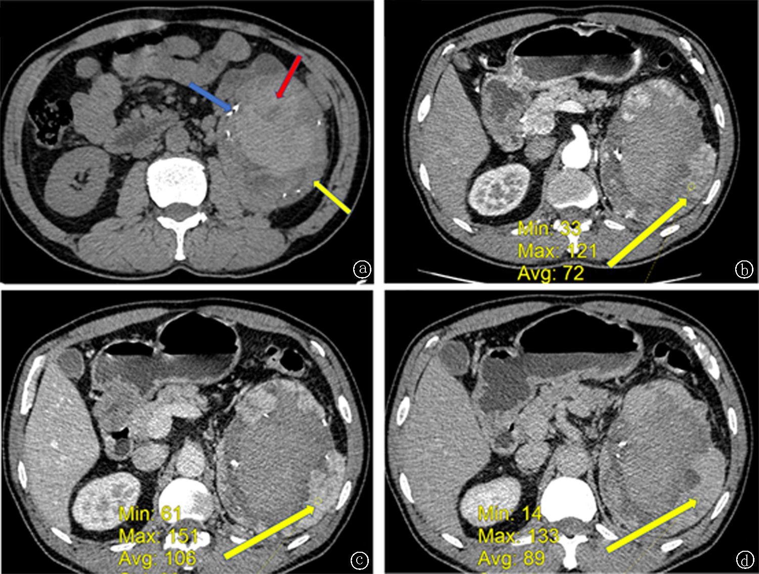
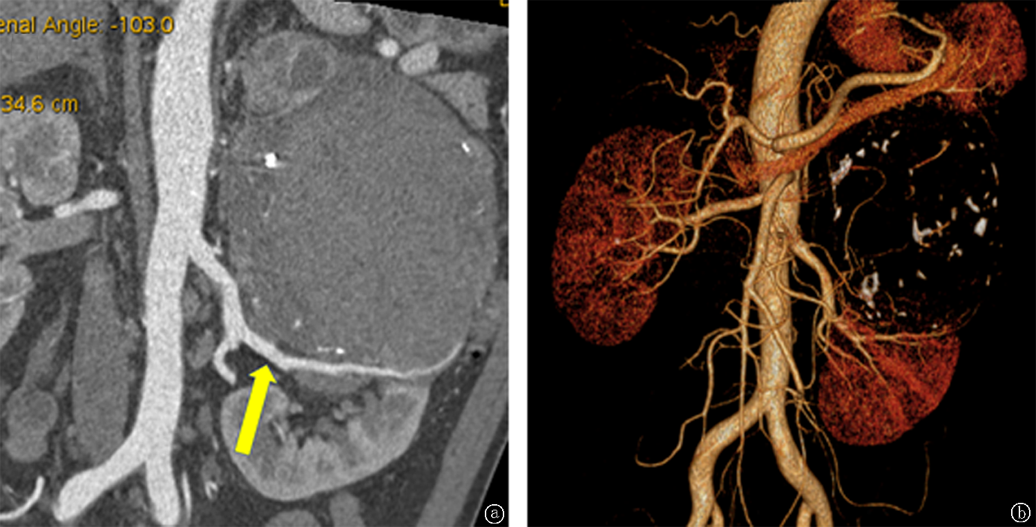
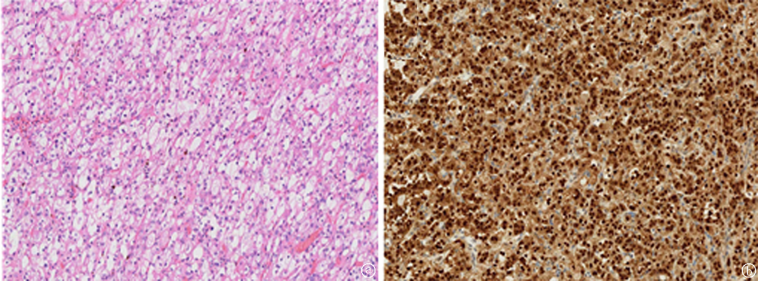
目的 报道1例TFEB易位肾细胞癌并进行文献复习,探讨TFEB易位肾细胞癌的临床及影像特点,以期提高对该病的认识。方法 回顾性分析1例TFEB易位肾细胞癌的临床及影像资料,并以中国知网、万方数据知识服务平台及PubMed 等为数据来源,通过“TFEB易位肾细胞癌”、“MiT家族易位性肾细胞癌”、“6p21 translocation renal cell carcinoma”以及“t (6;11)(p21;q12)”等关键词进行文献检索,筛选出从2010年1月以来公开发表的且有影像学资料的中英文文献,分析TFEB易位肾细胞癌的临床及影像特点。结果 本例为男性,54岁,腰痛伴肉眼血尿,CT平扫表现为边界清楚、囊实性肿块,伴出血及钙化,增强扫描呈不均匀强化,无远处转移。文献报道的13例确诊病例:男女比例为8∶5;平均年龄为37.8岁;肿块平均最大径约7.7 cm;均进行了手术切除,其中1例术前出现双肺转移,其余12例均无转移。影像学方面,肿块多边界清楚, 以囊实性混杂密度为主,强化方式以持续的轻中度强化为主。结论 TFEB易位肾细胞癌是一种少见的非侵袭性低度恶性肿瘤,发生于年轻患者、表现为边界清楚、轻中度强化的囊实性肿块,需要考虑到TFEB易位肾细胞癌的可能性。

关键词: 癌, 肾细胞, TFEB易位肾细胞癌, MiT家族易位性肾细胞癌

Abstract:

Objective To report a case of TFEB translocation renal cell carcinoma and review the literature, to explore its clinical and imaging features, in order to improve the understanding of the disease. Methods The clinical and imaging data of a case of TFEB translocation renal cell carcinoma were retrospectively analyzed, the “TFEB translocation renal cell carcinoma”, “MiT-family translocation renal cell carcinoma”, “6p21 translocation renal cell carcinoma”, and “t (6;11)(p21; q12)” were selected as keywords. The papers collected by China National Knowledge Internet (CNKI), Wanfang database and PubMed from January 2010 to present were retrieved. Clinical features and imaging characteristics of TFEB translocation renal cell carcinoma were summarized. Results A 54-year-old male patient was admitted to the hospital because of backache and gross hematuria. The computed tomography (CT) scan showed a cystic and solid mass with clear borders, accompanied by hemorrhage and calcification. The enhanced scan showed uneven enhancement and no distant metastasis. The 13 confirmed cases reported in the literature had a male to female ratio of 8∶5; the average age was 37.8 years; the average maximum diameter of the tumor was about 7.7 cm. The surgical resection of the tumor was performed in all cases, one case had bilateral lung metastasis before surgery, and the other 12 cases had no metastasis. The masses had clear boundaries, mainly cystic and solid mixed density, and the enhancement mode was mainly continuous mild to moderate enhancement. Conclusion TFEB translocation renal cell carcinoma is a rare non-invasive low-grade malignant tumor, which occurs in young patients with well-defined, mild to moderately enhanced cystic-solid masses, the possibility of TFEB translocation renal cell carcinoma needs to be considered.

Key words: carcinoma, renal cell, TFEB translocation renal cell carcinoma, MiT family translocation renal cell carcinoma

中图分类号: