临床荟萃 ›› 2023, Vol. 38 ›› Issue (10): 917-921.doi: 10.3969/j.issn.1004-583X.2023.10.010

• 论著 • 上一篇    下一篇

光学相干断层成像指导治疗变异型心绞痛1例并文献复习

王家琦1, 谢悦陶2, 高曼2, 宋学莲2, 张飞飞2, 党懿2, 齐晓勇2()   

  1. 1.河北北方学院研究生院, 河北 张家口 075000
    2.河北省人民医院 心血管内科,河北 石家庄 050051
  • 收稿日期:2022-07-16 出版日期:2023-10-20 发布日期:2024-01-03
  • 通讯作者: 齐晓勇 E-mail:hbghxiaoyong_q@126.com
  • 基金资助:
    河北省自然科学基金——基于TGFβ/Smad通路探讨心脏收缩力调节对心力衰竭心肌能量代谢的影响及机制(H2020307017);河北省2019年度医学科学研究课题——血管紧张素受体脑啡肽酶抑制剂对缺血性心肌损伤后心衰的防治研究(20190237)

Treatment of variant angina pectoris guided by the optical coherence tomography: A case report and literature review

Wang Jiaqi1, Xie Yuetao2, Gao Man2, Song Xuelian2, Zhang Feifei2, Dang Yi2, Qi Xiaoyong2()   

  1. 1. Graduate School of Hebei North University,Zhangjiakou 075000, China
    2. Department of Cardiology,Hebei General Hospital,Shijiazhuang 050051, China
  • Received:2022-07-16 Online:2023-10-20 Published:2024-01-03
  • Contact: Qi Xiaoyong E-mail:hbghxiaoyong_q@126.com

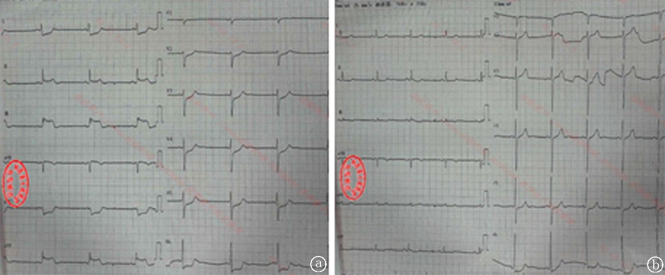
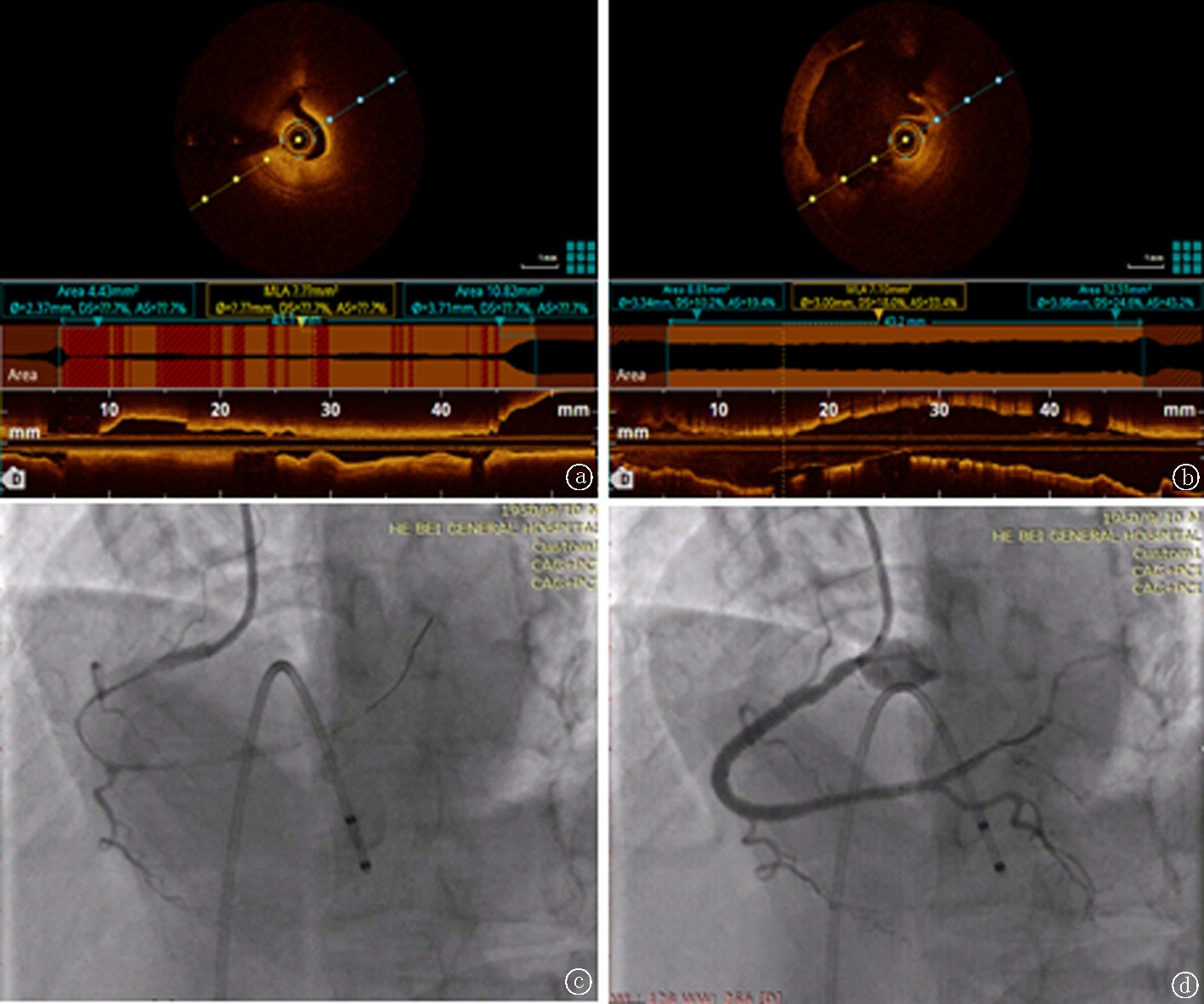
摘要: 目的 探讨变异型心绞痛的临床特点,以期提高对该病的诊疗与认识。方法 回顾性分析1例由光学相干断层成像指导治疗的变异型心绞痛患者临床资料,并以“变异型心绞痛”以及“光学相干断层成像”等关键词,通过检索中国知网、PubMed及万方数据库,筛选公开发表的中英文文献,以分析变异型心绞痛的临床特点。结果 本例患者老年男性,因间断胸闷2年余,加重10余天就诊,发作时心电图检查提示Ⅱ、Ⅲ、aVF导联ST段抬高,其余导联ST段显著压低,症状缓解后ST段回落,于光学相关断层成像指导下行冠状动脉造影结果示右冠状动脉近中段狭窄20%~30%,中段第二转折狭窄70%,远段可见支架影,右冠状动脉行麦角新碱激发试验,可见弥漫痉挛,给予硝酸甘油及硝普钠后痉挛解除,考虑患者胸闷症状反复发作,药物控制欠佳,且术中冠脉痉挛明显,遂给予右冠状动脉支架置入,经治疗患者胸闷症状未再发作,治疗效果良好。结论 变异型心绞痛疾病特点及治疗方法与冠状动脉粥样硬化不同,临床易误诊误治,因此提高对其的警惕性及认识,及早行腔内影像学检查及激发试验可明确诊断,有助于选择合适的治疗方案并改善患者预后。

关键词: 光学相干断层成像, 变异型心绞痛, 冠状动脉介入治疗

Abstract: Objective To explore the clinical characteristics of variant angina pectoris, thus improving its diagnosis and treatment. Methods The clinical data of a patient with variant angina pectoris who was treated under the guidance of optical coherence tomography (OCT) were retrospectively analyzed. The clinical features of variant angina pectoris were analyzed by searching relevant articles in the CNKI, PubMed and Wanfang database with key words of “variant angina pectoris” and “OCT”. Results The male older patient presented with intermittent chest for more than 2 years and aggravated for more than 10 days was admitted. Electrocardiography on attack showed ST elevations in leads of II, III and arteriovenous fistula (AVF), and downwards of the remaining leads. The ST segment fell back after symptoms were relieved. The results of coronary angiography under the guidance of OCT showed a stenosis of 20%-30% in the proximal and middle segments of the right coronary artery, a stenosis of 70% in the second turn of the middle segment, and a stent shadow in the distal segment. Ergonovine stimulation test in the right coronary artery showed diffuse spasm, which was relieved after the treatment of nitroglycerin and sodium nitroprusside. Considering the repeated attacks of chest tightness, poor response to medication andintraoperative coronary spasm, the patient was managed by stent implantationin the right coronary artery.After stent implantation, symptoms of chest tightness disappeared with a good efficacy. Conclusion Disease characteristics and treatment of variant angina differ from those of coronary atherosclerosis, presenting a high rate of misdiagnosis. It is necessary to improve the vigilance and understanding of the disease. Timelyintracavitary imaging examination and provocation test contribute to a precise diagnosis and favor the selection of the optimal treatment and improvement of the prognosis.

Key words: optical coherence tomography, variant angina pectoris, coronary intervention

中图分类号: