
临床荟萃 ›› 2023, Vol. 38 ›› Issue (12): 1067-1072.doi: 10.3969/j.issn.1004-583X.2023.12.002
陶嘉楠1, 李文茜2, 马秀雯2, 安琪1, 王学红1( )
)
收稿日期:2023-06-15
出版日期:2023-12-20
发布日期:2024-01-30
通讯作者:
王学红
E-mail:Lindawang0710@hotmail.com
基金资助:
Tao Jianan1, Li Wenqian2, Ma Xiuwen2, An Qi1, Wang Xuehong1()
Received:2023-06-15
Online:2023-12-20
Published:2024-01-30
Contact:
Wang Xuehong
E-mail:Lindawang0710@hotmail.com
摘要:
目的 系统评价人表皮生长因子受体-2(human epidermal growth factor receptor-2,HER-2)在肝细胞癌(hepatocellular carcinoma,HCC)中的表达及其与临床病理特征的关系。方法 计算机检索PubMed、中国知网、万方数据库和维普数据库,收集国内外关于HER-2在HCC中表达的病例-对照研究,检索时限为从建库至2023年5月。由2名研究者独立检索筛选文献、提取资料并评价偏倚风险,用Revman5.3软件进行meta分析。结果 最终纳入31个病例-对照研究,共计2558例患者。Meta分析结果显示:HER-2在HCC中的阳性率(54.3%)高于对照组(50.5%),差异具有统计学意义[
中图分类号:
陶嘉楠, 李文茜, 马秀雯, 安琪, 王学红. HER-2在肝细胞癌中表达及临床意义的meta分析[J]. 临床荟萃, 2023, 38(12): 1067-1072.
Tao Jianan, Li Wenqian, Ma Xiuwen, An Qi, Wang Xuehong. Meta-analysis of expression and clinical significance of HER-2 in hepatocellular carcinoma[J]. Clinical Focus, 2023, 38(12): 1067-1072.
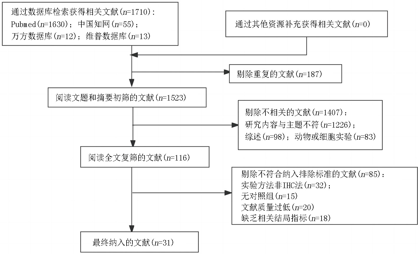
图1 文献筛选流程及结果
Fig.1 Process and results of Literature screening
| 序号 | 作者 | 发表时间 (年) | 国家 | 观察组 (例) | 性别 (男/女) | 平均年龄 (岁) | 观察组HER-2 阳性率(%) | 对照组HER-2 阳性率(%) | 结局指标 | NOS 评分 |
|---|---|---|---|---|---|---|---|---|---|---|
| 1 | Jiang等[ | 2018 | 中国 | 176 | 148/28 | 51.4±8.3(25-78) | 47.2(83/176) | - | b、d、e、f、g、h、i、j | 5 |
| 2 | Bassullu等[ | 2012 | 土耳其 | 50 | 44/6 | 56.8(26-72) | 92.0(46/50) | - | b、e、f、h、i | 5 |
| 3 | Bahnassi等[ | 2005 | 埃及 | 32 | 18/14 | 51.6(10-74) | 75(24/32) | 77.8(14/18) | a、e、f | 8 |
| 4 | Niu等[ | 2005 | 中国 | 41 | - | - | 24.4(10/41) | 12.9(4/31) | a | 8 |
| 5 | Xian等[ | 2005 | 中国 | 868 | 735/133 | 50.3(25-76) | 7.1(62/868) | - | b、d、j | 5 |
| 6 | Lin等[ | 2000 | 中国 | 50 | 34/16 | 50.1(18-72) | 70.0(35/50) | 22.0(11/50) | a | 7 |
| 7 | Ito等[ | 2001 | 日本 | 100 | 84/16 | 62.3±7.5 | 21.0(21/100) | - | d、e、h、i | 5 |
| 8 | Nakopoulou等[ | 1994 | 希腊 | 71 | 63/8 | 63.5(49-93) | 29.6(21/71) | 38.1(16/42) | a | 8 |
| 9 | Potti等[ | 2003 | 美国 | 25 | - | 66 | 8(2/25) | 0(0/25) | a | 6 |
| 10 | 李春丽等[ | 2013 | 中国 | 35 | 28/7 | 50.2(29-77) | 77.1(27/35) | 10.0(1/10) | a、b、e、f、h、i、j、k | 8 |
| 11 | 王东等[ | 2010 | 中国 | 127 | 112/15 | 51.7±12.6 | 22.8(29/127) | - | d、e、i、k | 5 |
| 12 | 刘换新等[ | 2007 | 中国 | 56 | 48/8 | 45.5(25-77) | 51.8(29/56) | 55.0(22/40) | a、b、c、h、j | 8 |
| 13 | 何怡等[ | 2005 | 中国 | 103 | 93/10 | 45.5(25-77) | 50.5(52/103) | 64.0(32/50) | a、b、c、j | 8 |
| 14 | 曾志武等[ | 2003 | 中国 | 26 | - | - | 38.5(10/26) | 5(1/20) | a | 8 |
| 15 | 姜建涛等[ | 2002 | 中国 | 32 | 27/5 | 48.0(22-70) | 75.0(24/32) | 42.9(9/21) | a、h、j、k | 8 |
| 16 | 魏红等[ | 2000 | 中国 | 30 | 23/7 | 42.6±10.7 | 60.0(18/30) | 91.3(21/23) | a、j | 8 |
| 17 | 申延琴等[ | 2000 | 中国 | 35 | - | - | 34.3(12/35) | 76.3(29/38) | a | 8 |
| 18 | 王影等[ | 1999 | 中国 | 45 | 42/3 | 48.5(25-72) | 55.6(25/45) | 28.9(13/45) | a、d | 7 |
| 19 | 牛兆山等[ | 1997 | 中国 | 41 | - | - | 24.4(10/41) | 16.7(3/18) | a | 8 |
| 20 | 陈晓江等[ | 1997 | 中国 | 28 | - | - | 53.6(15/28) | 42.9(12/28) | a | 7 |
| 21 | 周子成等[ | 1997 | 中国 | 46 | 41/5 | 47.3(23-64) | 47.8(22/46) | 44.7(17/38) | a | 8 |
| 22 | 李凡彩等[ | 1997 | 中国 | 41 | - | - | 26.8(11/41) | 22.0(9/41) | a、d | 7 |
| 23 | 张俊凯等[ | 2010 | 中国 | 30 | 19/11 | 56.8±9.6(38-85) | 40.0(12/30) | 10.0(1/10) | a、b、d、e、h、j、k | 8 |
| 24 | 李君达等[ | 1999 | 中国 | 60 | 45/15 | 50(25-74) | 31.7(19/60) | 3.7(1/27) | a、d | 8 |
| 25 | 程瑞雪等[ | 1997 | 中国 | 40 | - | - | 80(32/40) | 52.5(21/40) | a | 7 |
| 26 | 洪丰等[ | 1997 | 中国 | 55 | - | - | 52.7(29/55) | 71.7(27/38) | a | 8 |
| 27 | 王淼等[ | 1997 | 中国 | 68 | - | - | 100(68/68) | 77.1(74/96) | a | 8 |
| 28 | 王万忠等[ | 1996 | 中国 | 76 | 49/27 | 51.3(26-76) | 57.9(44/76) | 71.9(41/57) | a、j | 8 |
| 29 | 苏勤等[ | 1995 | 中国 | 55 | - | - | 100(52/52) | 81.0(111/137) | a | 8 |
| 30 | 张迎等[ | 2017 | 中国 | 82 | 53/29 | 45.4±6.8 | 59.8(49/82) | - | d,g,h | 5 |
| 31 | 林国跃等[ | 1998 | 中国 | 34 | - | - | 64.7(22/34) | 22.0(11/50) | a | 8 |
表1 纳入研究的基本特征及偏倚风险评价结果
Tab.1 Basic features and risk of bias assessment of included studies
| 序号 | 作者 | 发表时间 (年) | 国家 | 观察组 (例) | 性别 (男/女) | 平均年龄 (岁) | 观察组HER-2 阳性率(%) | 对照组HER-2 阳性率(%) | 结局指标 | NOS 评分 |
|---|---|---|---|---|---|---|---|---|---|---|
| 1 | Jiang等[ | 2018 | 中国 | 176 | 148/28 | 51.4±8.3(25-78) | 47.2(83/176) | - | b、d、e、f、g、h、i、j | 5 |
| 2 | Bassullu等[ | 2012 | 土耳其 | 50 | 44/6 | 56.8(26-72) | 92.0(46/50) | - | b、e、f、h、i | 5 |
| 3 | Bahnassi等[ | 2005 | 埃及 | 32 | 18/14 | 51.6(10-74) | 75(24/32) | 77.8(14/18) | a、e、f | 8 |
| 4 | Niu等[ | 2005 | 中国 | 41 | - | - | 24.4(10/41) | 12.9(4/31) | a | 8 |
| 5 | Xian等[ | 2005 | 中国 | 868 | 735/133 | 50.3(25-76) | 7.1(62/868) | - | b、d、j | 5 |
| 6 | Lin等[ | 2000 | 中国 | 50 | 34/16 | 50.1(18-72) | 70.0(35/50) | 22.0(11/50) | a | 7 |
| 7 | Ito等[ | 2001 | 日本 | 100 | 84/16 | 62.3±7.5 | 21.0(21/100) | - | d、e、h、i | 5 |
| 8 | Nakopoulou等[ | 1994 | 希腊 | 71 | 63/8 | 63.5(49-93) | 29.6(21/71) | 38.1(16/42) | a | 8 |
| 9 | Potti等[ | 2003 | 美国 | 25 | - | 66 | 8(2/25) | 0(0/25) | a | 6 |
| 10 | 李春丽等[ | 2013 | 中国 | 35 | 28/7 | 50.2(29-77) | 77.1(27/35) | 10.0(1/10) | a、b、e、f、h、i、j、k | 8 |
| 11 | 王东等[ | 2010 | 中国 | 127 | 112/15 | 51.7±12.6 | 22.8(29/127) | - | d、e、i、k | 5 |
| 12 | 刘换新等[ | 2007 | 中国 | 56 | 48/8 | 45.5(25-77) | 51.8(29/56) | 55.0(22/40) | a、b、c、h、j | 8 |
| 13 | 何怡等[ | 2005 | 中国 | 103 | 93/10 | 45.5(25-77) | 50.5(52/103) | 64.0(32/50) | a、b、c、j | 8 |
| 14 | 曾志武等[ | 2003 | 中国 | 26 | - | - | 38.5(10/26) | 5(1/20) | a | 8 |
| 15 | 姜建涛等[ | 2002 | 中国 | 32 | 27/5 | 48.0(22-70) | 75.0(24/32) | 42.9(9/21) | a、h、j、k | 8 |
| 16 | 魏红等[ | 2000 | 中国 | 30 | 23/7 | 42.6±10.7 | 60.0(18/30) | 91.3(21/23) | a、j | 8 |
| 17 | 申延琴等[ | 2000 | 中国 | 35 | - | - | 34.3(12/35) | 76.3(29/38) | a | 8 |
| 18 | 王影等[ | 1999 | 中国 | 45 | 42/3 | 48.5(25-72) | 55.6(25/45) | 28.9(13/45) | a、d | 7 |
| 19 | 牛兆山等[ | 1997 | 中国 | 41 | - | - | 24.4(10/41) | 16.7(3/18) | a | 8 |
| 20 | 陈晓江等[ | 1997 | 中国 | 28 | - | - | 53.6(15/28) | 42.9(12/28) | a | 7 |
| 21 | 周子成等[ | 1997 | 中国 | 46 | 41/5 | 47.3(23-64) | 47.8(22/46) | 44.7(17/38) | a | 8 |
| 22 | 李凡彩等[ | 1997 | 中国 | 41 | - | - | 26.8(11/41) | 22.0(9/41) | a、d | 7 |
| 23 | 张俊凯等[ | 2010 | 中国 | 30 | 19/11 | 56.8±9.6(38-85) | 40.0(12/30) | 10.0(1/10) | a、b、d、e、h、j、k | 8 |
| 24 | 李君达等[ | 1999 | 中国 | 60 | 45/15 | 50(25-74) | 31.7(19/60) | 3.7(1/27) | a、d | 8 |
| 25 | 程瑞雪等[ | 1997 | 中国 | 40 | - | - | 80(32/40) | 52.5(21/40) | a | 7 |
| 26 | 洪丰等[ | 1997 | 中国 | 55 | - | - | 52.7(29/55) | 71.7(27/38) | a | 8 |
| 27 | 王淼等[ | 1997 | 中国 | 68 | - | - | 100(68/68) | 77.1(74/96) | a | 8 |
| 28 | 王万忠等[ | 1996 | 中国 | 76 | 49/27 | 51.3(26-76) | 57.9(44/76) | 71.9(41/57) | a、j | 8 |
| 29 | 苏勤等[ | 1995 | 中国 | 55 | - | - | 100(52/52) | 81.0(111/137) | a | 8 |
| 30 | 张迎等[ | 2017 | 中国 | 82 | 53/29 | 45.4±6.8 | 59.8(49/82) | - | d,g,h | 5 |
| 31 | 林国跃等[ | 1998 | 中国 | 34 | - | - | 64.7(22/34) | 22.0(11/50) | a | 8 |
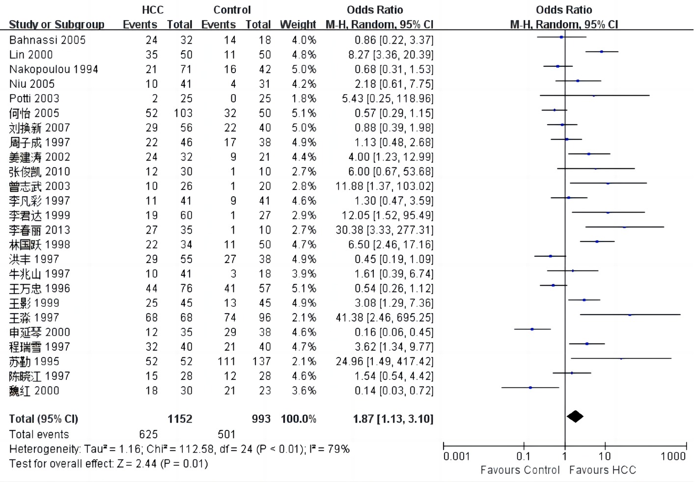
图2 HER-2在HCC中表达情况的meta分析
Fig.2 Meta-analysis of HER-2 expression in HCC
| 临床病理特征 | 纳入研究数 | 异质性检验结果 | 效应模型 | meta分析结果 | ||
|---|---|---|---|---|---|---|
| 性别(男/女) | 7[ | 0 | 0.99 | 固定 | 1.22(0.79,1.91) | 0.37 |
| 年龄(≥49岁/<49岁组) | 2[ | 0 | 0.96 | 固定 | 0.48(0.25,0.94) | 0.03 |
| 分化程度(低分化/中-高分化) | 9[ | 39 | 0.11 | 固定 | 2.61(1.84,3.69) | <0.01 |
| 肿瘤直径(≥5 cm/<5 cm) | 7[ | 14 | 0.32 | 固定 | 1.93(1.25,2.98) | 0.003 |
| 淋巴结转移(有/无) | 4[ | 52 | 0.10 | 随机 | 3.24(0.69,15.31) | 0.14 |
| 血行转移(有/无) | 2[ | 0 | 0.82 | 固定 | 5.42(3.15,9.31) | <0.01 |
| 临床分期(Ⅲ、Ⅳ期/Ⅰ、Ⅱ期) | 8[ | 39 | 0.12 | 固定 | 2.02(1.39,2.94) | 0.0003 |
| 门静脉侵犯(有/无) | 5[ | 42 | 0.14 | 固定 | 1.73(1.11,2.72) | 0.02 |
| 血清HBsAg(+/-) | 9[ | 33 | 0.16 | 固定 | 2.22(1.55,3.18) | <0.01 |
| 血清AFP(≥400 μg/L组/<400 μg/L) | 4[ | 0 | 0.45 | 固定 | 1.02(0.55,1.88) | 0.95 |
表2 HER-2在HCC中表达与临床病理特征的meta分析
Tab.2 Meta-analysis of the relationship between the expression of HER-2 and clinicopathologic features in HCC
| 临床病理特征 | 纳入研究数 | 异质性检验结果 | 效应模型 | meta分析结果 | ||
|---|---|---|---|---|---|---|
| 性别(男/女) | 7[ | 0 | 0.99 | 固定 | 1.22(0.79,1.91) | 0.37 |
| 年龄(≥49岁/<49岁组) | 2[ | 0 | 0.96 | 固定 | 0.48(0.25,0.94) | 0.03 |
| 分化程度(低分化/中-高分化) | 9[ | 39 | 0.11 | 固定 | 2.61(1.84,3.69) | <0.01 |
| 肿瘤直径(≥5 cm/<5 cm) | 7[ | 14 | 0.32 | 固定 | 1.93(1.25,2.98) | 0.003 |
| 淋巴结转移(有/无) | 4[ | 52 | 0.10 | 随机 | 3.24(0.69,15.31) | 0.14 |
| 血行转移(有/无) | 2[ | 0 | 0.82 | 固定 | 5.42(3.15,9.31) | <0.01 |
| 临床分期(Ⅲ、Ⅳ期/Ⅰ、Ⅱ期) | 8[ | 39 | 0.12 | 固定 | 2.02(1.39,2.94) | 0.0003 |
| 门静脉侵犯(有/无) | 5[ | 42 | 0.14 | 固定 | 1.73(1.11,2.72) | 0.02 |
| 血清HBsAg(+/-) | 9[ | 33 | 0.16 | 固定 | 2.22(1.55,3.18) | <0.01 |
| 血清AFP(≥400 μg/L组/<400 μg/L) | 4[ | 0 | 0.45 | 固定 | 1.02(0.55,1.88) | 0.95 |
| [1] |
Forner A, Reig M, Bruix J. Hepatocellular carcinoma[J]. Lancet, 2018, 391(10127):1301-1314.
doi: S0140-6736(18)30010-2 pmid: 29307467 |
| [2] |
Bray F, Ferlay J, Soerjomataram I, et al. Global cancer statistics 2018: GLOBOCAN estimates of incidence and mortality worldwide for 36 cancers in 185 countries[J]. CA Cancer J Clin, 2018, 68(6):394-424.
doi: 10.3322/caac.v68.6 URL |
| [3] | 国家卫生健康委办公厅. 原发性肝癌诊疗指南(2022年版)[J]. 中华外科杂志, 2022, 60(4):273-309. |
| [4] | de Abreu Pereira D, Sandim V, Fernandes T, et al. Proteomic analysis of HCC-1954 and MCF-7 cell lines highlights crosstalk between αv and β1 integrins, E-cadherin and HER-2[J]. Int J MolSci,2022, 23(17):10194. |
| [5] |
Owis AI, Sherif NH, Hassan AA, et al. Tropaeolum majus L. and low dose gamma radiation suppress liver carcinoma development via EGFR-HER2 signaling pathway[J]. Nat Prod Res, 2023, 37(6):1030-1035.
doi: 10.1080/14786419.2022.2098958 URL |
| [6] | Chen G, Jiang J, Wang X, et al. lncENSTsuppress the warburg effect regulating the tumor progress by the Nkx2-5/ErbB2 axis in hepatocellular carcinoma[J]. Comput Math Methods Med, 2021:6959557. |
| [7] |
Loibl S, Poortmans P, Morrow M, et al. Breast cancer[J]. Lancet, 2021, 397(10286):1750-1769.
doi: 10.1016/S0140-6736(20)32381-3 pmid: 33812473 |
| [8] | 吕晓双, 罗斌. 抗HER-2治疗的耐药机制及对策的研究进展[J]. 现代肿瘤医学, 2022, 30(3):529-534. |
| [9] | 陶嘉楠, 田王钊, 安琪, 等. SLP-2在胃癌中的表达及临床意义的Meta分析[J]. 临床荟萃, 2022, 37(4):299-304. |
| [10] |
Jiang LH, Hao YL, Zhu JW. Expression and prognostic value of HER-2/neu, STAT3 and SOCS3 in hepatocellular carcinoma[J]. Clin Res Hepatol Gastroenterol, 2019, 43(3):282-291.
doi: 10.1016/j.clinre.2018.09.011 URL |
| [11] | Bassullu N, Turkmen I, Dayangac M, et al. The predictive and prognostic significance of c-erb-B2, EGFR, PTEN, mTOR, PI3K, p27, and ERCC1 expression in hepatocellular carcinoma[J]. Hepat Mon, 2012, 12(10 HCC):e7492. |
| [12] |
Bahnassi AA, Zekri AR, El-Houssini S, et al. Hepatitis C virus-NS3P in relation to p53, p21waf, mdm2, p21-ras and c-erbB2 in hepatocarcinogenesis[J]. J Gastroenterol Hepatol, 2005, 20(11):1731-1740.
doi: 10.1111/jgh.2005.20.issue-11 URL |
| [13] |
Niu ZS, Wang M. Expression of c-erbB-2 and glutathione S-transferase-pi in hepatocellular carcinoma and its adjacent tissue[J]. World J Gastroenterol, 2005, 11(28):4404-4408.
doi: 10.3748/wjg.v11.i28.4404 URL |
| [14] |
Xian ZH, Zhang SH, Cong WM, et al. Overexpression/amplification of HER-2/neu is uncommon in hepatocellular carcinoma[J]. J Clin Pathol, 2005, 58(5):500-503.
doi: 10.1136/jcp.2004.023556 pmid: 15858121 |
| [15] | Lin GY, Chen ZL, Lu CM, et al. Immunohistochemical study on p53, H-rasp21, c-erbB-2 protein and PCNA expression in HCC tissues of Han and minority ethnic patients[J]. World J Gastroenterol, 2000, 6(2):234-238. |
| [16] |
Ito Y, Takeda T, Sakon M, et al. Expression and clinical significance of erb-B receptor family in hepatocellular carcinoma[J]. Br J Cancer, 2001, 84(10):1377-1383.
doi: 10.1054/bjoc.2000.1580 URL |
| [17] |
Nakopoulou L, Stefanaki K, Filaktopoulos D, et al. C-erb-B-2 oncoprotein and epidermal growth factor receptor in human hepatocellular carcinoma: an immunohistochemical study[J]. Histol Histopathol, 1994, 9(4):677-682.
pmid: 7894139 |
| [18] |
Potti A, Ganti AK, Tendulkar K, et al. HER-2/neu and CD117 (C-kit) overexpression in hepatocellular and pancreatic carcinoma[J]. Anticancer Res, 2003, 23(3B):2671-2674.
pmid: 12894556 |
| [19] | 李春丽, 田涛, 南克俊. c-erbB-2、ER及PR在肝癌组织中的表达及临床意义[J]. 现代肿瘤医学, 2013, 21(9):2029-2032. |
| [20] | 王东, 朱继业, 栗光明, 等. CD117、C-erbB-2及表皮生长因子受体在原发性肝癌组织中的表达及其临床意义[J]. 中华实验外科杂志, 2010, 27(12):1815-1817. |
| [21] | 刘换新, 杨磊, 袁怀文, 等. CD117、C-erbB-2及Ki-67蛋白在原发性肝细胞性肝癌中的表达及意义[J]. 肝胆外科杂志, 2007, 15(3):228-231. |
| [22] | 何怡, 王东, 李增鹏, 等. CD117、C-erbB-2蛋白在原发性肝细胞性肝癌中的表达及意义[J]. 消化外科, 2005, 4(2):103-107. |
| [23] | 曾志武, 易继林, 刘天威, 等. HER-2/neu基因在原发性肝癌中的表达及意义[J]. 临床外科杂志, 2003, 11(5):282-284. |
| [24] | 姜建涛, 李国威, 赵敏, 等. EGFR、C-ErbB-2蛋白在原发性肝细胞性肝癌中的表达及意义[J]. 中国肿瘤临床与康复, 2002, 9(3):9-11. |
| [25] | 魏红, 李永国, 杨竹林. 原发性肝癌和慢性肝病组织中c-erbB-2、TGF-α的表达及意义[J]. 肝胆胰外科杂志, 2000, 12(4):178-179. |
| [26] | 申延琴, 黄致治. 癌基因c-erbB-2蛋白在肝细胞癌中的表达(英文)[J]. 汕头大学医学院学报, 2000, 13(3):3-4,12. |
| [27] | 王影, 杨素琼, 王在国, 等. 原发性肝细胞癌中ras P21, C-erbB-2和P16蛋白的表达意义[J]. 世界华人消化杂志, 1999, 7(9):808-809. |
| [28] | 牛兆山, 张昭成, 邹伟. 人肝癌及癌旁组织中原癌基因c-erbB-2蛋白产物的表达[J]. 齐鲁肿瘤杂志, 1997, 4(4):23-24. |
| [29] | 陈晓江, 张漫凌, 王福民. C-erbB-2和P_(53)蛋白在肝癌中的表达的免疫组化研究[J]. 齐齐哈尔医学院学报, 1997, 18(4):246-248. |
| [30] | 周子成, 汪荣泉, 杨建民, 等. 肝癌患者HCV感染与癌基因c-erbB-2表达的关系[J]. 第三军医大学学报, 1997, 19(3):82-83. |
| [31] | 李凡彩, 曾思恩, 浣孝强, 等. c-erbB-2 和 p53 在小肝癌的表达与预后关系[J]. 中华外科杂志, 1997, 35(5):33. |
| [32] | 张俊凯, 潘佩玲, 吴颖猛, 等. HER-2/neu基因在肝细胞癌中的表达及意义[J]. 南方医科大学学报, 2010, 30(2):326-328. |
| [33] | 李君达, 王先法, 杨进, 等. 肝细胞癌癌基因P53, P21, C-erbB-2及EGFR表达的研究[J]. 齐齐哈尔医学院学报, 1999, 20(2):96-98. |
| [34] | 程瑞雪, 冯德云, 沈明, 等. c-erbB-2蛋白和HBsAg在肝细胞癌与癌旁组织中的表达及相互关系[J]. 中华病理学杂志, 1997, 26(5):308. |
| [35] | 洪丰, 白守国, 王万忠, 等. PCNA、C-erbB-2P-(185)在 HBV 感染慢性肝炎 LC、HCC 中表达的对照研究[J]. 济宁医学院学报, 1997, 20(2):15-17. |
| [36] | 王淼, 施公胜, 池鸣鸣, 等. 表皮生长因子受体和 C-erbB-2 蛋白在人肝细胞癌中的表达[J]. 南通医学院学报, 1997, 17(2):29-30. |
| [37] | 王万忠, 杜德利, 王家耀, 等. 肝细胞癌及癌旁组织中c-erbB-2蛋白的表达与乙型肝炎病毒感染的关系[J]. 中华肝脏病杂志, 1996, 4(3):139-141. |
| [38] | 苏勤, 刘彦仿. c-erbB-2蛋白和表皮生长因子受体在肝脏病变中的表达[J]. 中华病理学杂志, 1995, 24(2):93-95. |
| [39] | 张迎, 徐瀚峰, 万莉, 等. LOXL2与HER-2蛋白在原发性肝癌中的表达及其临床意义[J]. 广西医科大学学报, 2017, 34(8):1189-1191. |
| [40] | 林国跃, 陈朝伦, 吕才模, 等. c-erbB-2、H-ras p21、p53及PCNA在原发性肝癌中的表达[J]. 实用癌症杂志, 1998, 13(2):19-22. |
| [41] |
Chupradit S, Jasim SA, Bokov D, et al. Recent advances in biosensor devices for HER-2 cancer biomarker detection[J]. Anal Methods, 2022, 14(13):1301-1310.
doi: 10.1039/D2AY00111J URL |
| [42] |
Krishnamurti U, Silverman JF. HER2 in breast cancer: A review and update[J]. Adv Anat Pathol, 2014, 21(2):100-107.
doi: 10.1097/PAP.0000000000000015 pmid: 24508693 |
| [43] |
Fourati N, Trigui R, Charfeddine S, et al. [Concomitant radiotherapy and trastuzumab: Rational and clinical implications][J]. Bull Cancer, 2021, 108(5):501-512.
doi: 10.1016/j.bulcan.2020.12.012 pmid: 33745737 |
| [44] |
Palle J, Rochand A, Pernot S, et al. Human epidermal growth factor receptor 2 (HER2) in advanced gastric cancer: Current knowledge and future perspectives[J]. Drugs, 2020, 80(4):401-415.
doi: 10.1007/s40265-020-01272-5 pmid: 32077003 |
| [45] | 林莉, 王尤, 周福祥. 胃癌组织HER-2表达Meta分析[J]. 中华肿瘤防治杂志, 2015, 22(23):1843-1847. |
| [46] |
Cronin KA, Harlan LC, Dodd KW, et al. Population-based estimate of the prevalence of HER-2 positive breast cancer tumors for early stage patients in the US[J]. Cancer Invest, 2010, 28(9):963-968.
doi: 10.3109/07357907.2010.496759 pmid: 20690807 |
| [47] |
Yuan Y, Liu X, Cai Y, et al. Lapatinib and lapatinib plus trastuzumab therapy versus trastuzumab therapy for HER2 positive breast cancer patients: an updated systematic review and meta-analysis[J]. Syst Rev, 2022, 11(1):264.
doi: 10.1186/s13643-022-02134-9 pmid: 36496473 |
| [1] | 龚财芳, 赵俊宇, 游川. 接纳与承诺疗法对癌症患者心理健康和生活质量影响的meta分析[J]. 临床荟萃, 2024, 39(2): 101-107. |
| [2] | 肖煌怡, 袁建坤, 严梓予, 曾雯姝, 鲁兰莫, 王峻. 认知干预对遗忘型轻度认知障碍老年患者干预效果的meta分析[J]. 临床荟萃, 2024, 39(1): 12-19. |
| [3] | 吕畅, 周利明. TNF-α-308基因多态性与胃癌易感相关性的meta分析[J]. 临床荟萃, 2023, 38(9): 779-787. |
| [4] | 李海, 刘文虎, 彭绍鹏, 王飞. 控制性阶梯式减压术对比快速标准大骨瓣减压术治疗重度颅脑损伤疗效的meta分析[J]. 临床荟萃, 2023, 38(9): 788-795. |
| [5] | 侯有玲, 李奕, 关红玉, 罗红霞. 目标导向液体治疗在脑肿瘤切除术中应用效果的meta分析[J]. 临床荟萃, 2023, 38(8): 686-693. |
| [6] | 金家辉, 杨阳, 秦铜, 何雨欣, 苏美华. 补充益生菌对2型糖尿病患者糖代谢改善的meta分析[J]. 临床荟萃, 2023, 38(7): 581-587. |
| [7] | 肖王静, 李欣梦, 卢松玲, 孙雪华. 重复经颅磁刺激治疗中枢神经源性吞咽障碍疗效及安全性的meta分析[J]. 临床荟萃, 2023, 38(7): 588-599. |
| [8] | 尤奕, 高淑清, 徐浩. 肠内营养对食管癌患者术后临床结局影响的系统综述[J]. 临床荟萃, 2023, 38(6): 485-492. |
| [9] | 倪艺芸, 刘彬, 梁琪, 李晓凤. 白细胞介素6和C反应蛋白预测新型冠状病毒肺炎严重程度的meta分析[J]. 临床荟萃, 2023, 38(6): 493-499. |
| [10] | 沃拉孜汗·玛德尼亚提, 迪力夏提·图尔迪麦麦提, 李梦晨, 拜合提尼沙·吐尔地. 宏基因组二代测序技术在肺结核诊断中应用价值的meta分析[J]. 临床荟萃, 2023, 38(5): 389-398. |
| [11] | 赵哲, 穆培娟, 张冬. 恩度联合顺铂胸腔灌注治疗肺癌合并恶性胸腔积液疗效的meta分析[J]. 临床荟萃, 2023, 38(5): 399-404. |
| [12] | 马明福, 魏志国, 何铁英. 急性胰腺炎并发胰腺假性囊肿危险因素的meta分析[J]. 临床荟萃, 2023, 38(4): 293-301. |
| [13] | 曹宇萌, 张海燕, 刘立新. 非酒精性脂肪性肝病的病理改变与血清铁蛋白和血清铁含量变化关系的meta分析[J]. 临床荟萃, 2023, 38(3): 197-207. |
| [14] | 马宏莉, 陆皓, 王丹, 焦海星, 李一珂, 李思雨, 吕静. 脑卒中患者残疾危险因素的meta分析[J]. 临床荟萃, 2023, 38(2): 111-116. |
| [15] | 柯孟婷, 陈慰. 瑞舒伐他汀降压作用的meta分析[J]. 临床荟萃, 2023, 38(11): 965-971. |
| 阅读次数 | ||||||
|
全文 |
|
|||||
|
摘要 |
|
|||||