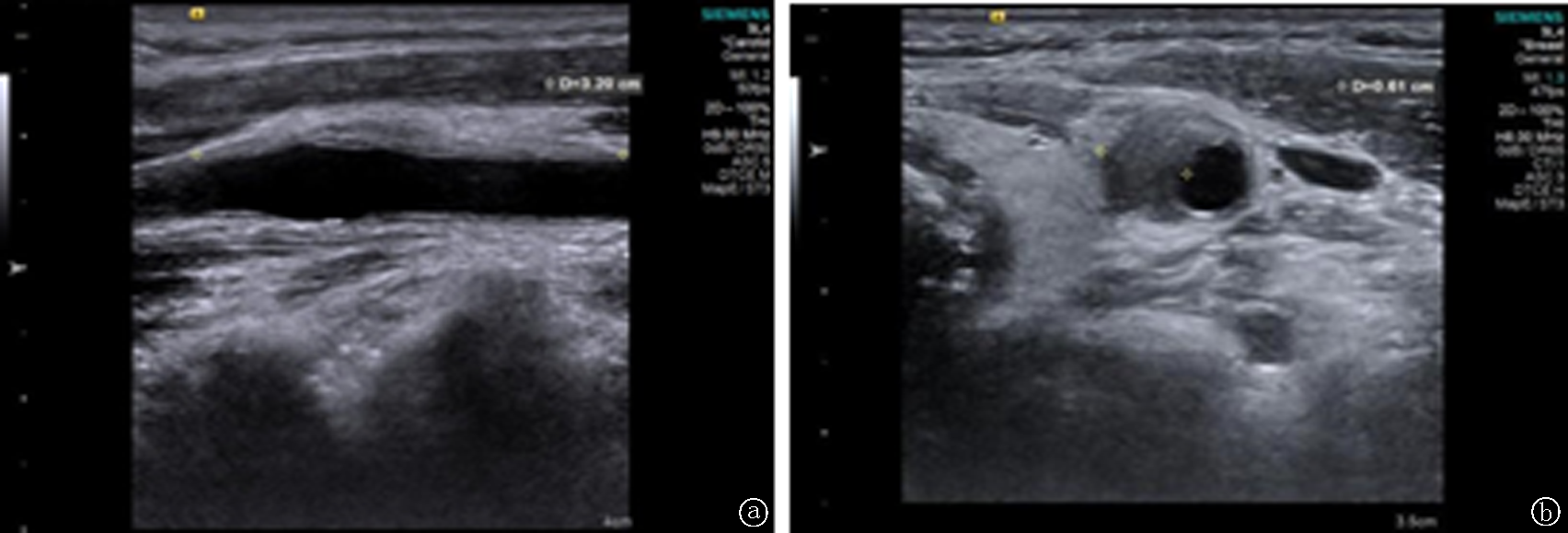
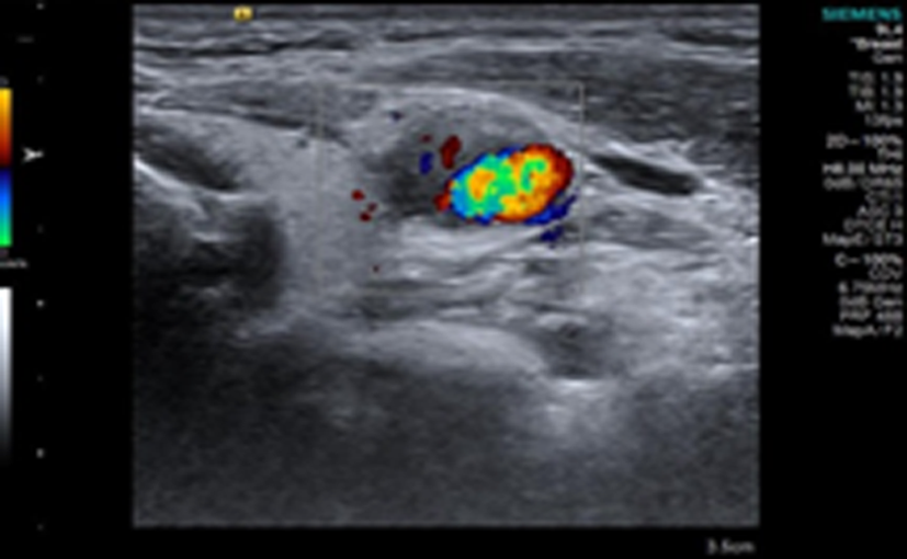
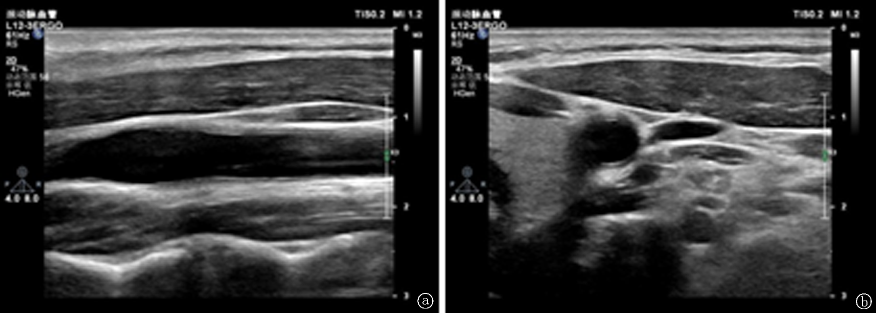
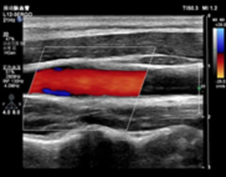
| [1] |
Coulier B, Van den Broeck S, Colin GC. Carotidynia alias transient perivascular inflammation of the carotid artery (TIPIC syndrome)[J]. J Belg Soc Radiol, 2018, 102(1):50.
doi: 10.5334/jbsr.1595
pmid: 30094415
|
| [2] |
Lecler A, Obadia M, Savatovsky J, et al. TIPIC syndrome: Beyond the myth of carotidynia, a new distinct unclassified entity[J]. AJNR Am J Neuroradiol, 2017, 38(7):1391-1398.
doi: 10.3174/ajnr.A5214
URL
|
| [3] |
栾艳艳, 王玉玲, 王春光. 短暂性颈动脉血管周围炎症综合征超声表现1例[J]. 中国超声医学杂志, 2022, 38(4):478.
|
| [4] |
费正东, 王磊, 王红慧, 等. 超声诊断短暂颈动脉周围炎症综合征1例[J]. 中华超声影像学杂志, 2019, 28(8):726-727.
|
| [5] |
张明珠, 高榆秀, 张玉竹, 等. 短暂颈动脉周围炎症综合征超声表现1例[J]. 中国临床医学影像杂志, 2022, 33(3):225-226.
|
| [6] |
Fay T. Atypical neuralgia[J]. Arch Neurol Psychiatry, 1927, 18:309-315.
|
| [7] |
Headache Classification Committee of the International Headache Society. Classification and diagnostic criteria for headache disorders, cranial neuralgias and facial pain[J]. Cepalalgia, 1988, 8(Suppl 7):1.
|
| [8] |
Headache Classification Committee of the International Headache Society. Classification and diagnostic criteria for headache disorders, cranial neuralgias, and facial pain[J]. Cephalalgia, 2004, 24(Suppl 1):1.
|
| [9] |
Rafailidis V, Chryssogonidis I, Tegos T, et al. Role of multi-parametric ultrasound in transient perivascular inflammation of the carotid artery syndrome[J]. Ultrasound, 2019, 27(2):77-84.
doi: 10.1177/1742271X18822658
pmid: 31037091
|
| [10] |
杨中华. AJNR:短暂颈动脉周围炎症综合征[J]. 中国卒中杂志, 2018, 13(3):272-273.
|
| [11] |
金晖, 刘尚全. 2型糖尿病患者血清胆红素水平与颈动脉斑块的相关性[J]. 临床荟萃, 2021, 36(4):340-343.
|
| [12] |
邓余杰, 范林清, 张涛. 脂蛋白相关磷脂酶A2与中老年高血压患者颈动脉不稳定斑块的关系[J]. 临床荟萃, 2021, 36(2):112-116.
|
| [13] |
赵梦婷, 王红春, 石燕清, 等. 颈动脉斑块积分及炎性因子与脑梗死神经功能缺损的相关性[J]. 临床荟萃, 2017, 32(6):491-494.
|
| [14] |
陈玮, 鄢磊, 阮琴韵, 等. 颈动脉夹层的超声表现及漏诊分析[J]. 中国医学影像技术, 2019, 35(11):1643-1647.
|
| [15] |
喻晓娜, 任卫东. 大动脉炎多模态血管超声的研究进展[J]. 医学综述, 2020, 26(3):540-543, 548.
|
 )
)