Clinical Focus ›› 2024, Vol. 39 ›› Issue (2): 108-114.doi: 10.3969/j.issn.1004-583X.2024.02.002
Previous Articles Next Articles
Diagnostic of superb microvascular imaging for breast tumors: A meta-analysis
Chen Sihan, Xiong Hu( ), Shui Dianya, Gao Xiaozhan, Liu Zewei
- Department of Ultrasound, the Second People's Hospital of Yichang, Yichang 443001, China
-
Received:2023-08-20Online:2024-02-20Published:2024-04-18 -
Contact:Xiong Hu E-mail:106722251@qq.com
CLC Number:
Cite this article
Chen Sihan, Xiong Hu, Shui Dianya, Gao Xiaozhan, Liu Zewei. Diagnostic of superb microvascular imaging for breast tumors: A meta-analysis[J]. Clinical Focus, 2024, 39(2): 108-114.
share this article
Add to citation manager EndNote|Ris|BibTeX
URL: https://huicui.hebmu.edu.cn/EN/10.3969/j.issn.1004-583X.2024.02.002
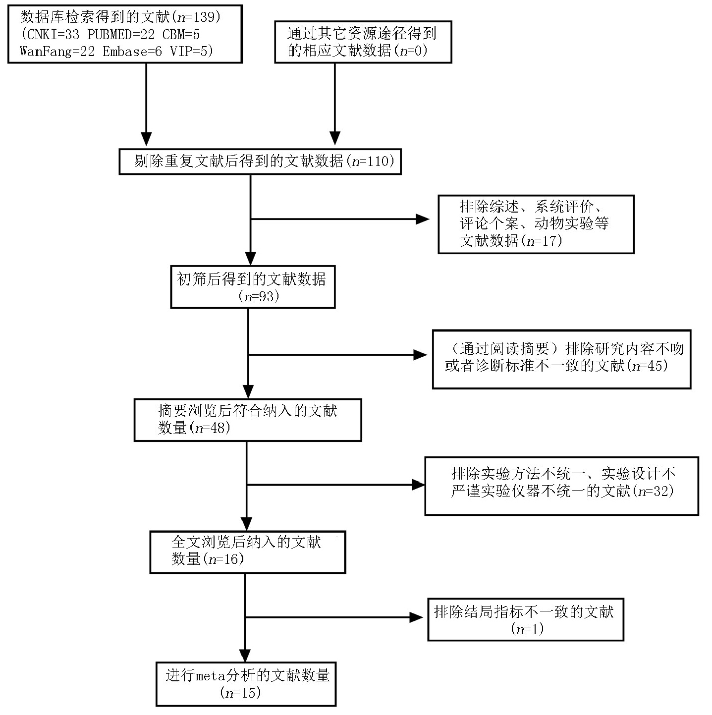
Fig.1 Flowchart for inclusion of primary literature
| 纳入研究 | 研究类型 | 超声仪器 | 病人数 | 纳入总数 (恶性/良性)(例) | 患者年龄 (岁) | TP (例) | FP (例) | FN (例) | TN (例) |
|---|---|---|---|---|---|---|---|---|---|
| Yongfeng[ | 回顾性 | TOSHIBA Aplio500 | 111 | 135(41/94) | 18~65 | 40 | 18 | 1 | 76 |
| Zhang[ | 回顾性 | TOSHIBA Aplio500 | 232 | 236(121/115) | 19~65 | 103 | 8 | 18 | 107 |
| Zhu[ | 回顾性 | TOSHIBA Aplio500 | 95 | 95(78/12) | 22~73 | 78 | 2 | 8 | 12 |
| Kim[ | 回顾性 | TOSHIBA Aplio500 | 65 | 62(12/50) | 28~80 | 10 | 6 | 2 | 44 |
| Xiao[ | 回顾性 | TOSHIBA Aplio400 | 132 | 132(58/74) | 16~78 | 45 | 7 | 13 | 67 |
| Cai[ | 回顾性 | TOSHIBA Aplio500 | 224 | 238(138/100) | NA | 64 | 4 | 48 | 109 |
| 张剑[ | 回顾性 | TOSHIBA Aplio400 | 85 | 87(41/46) | 33~80 | 30 | 12 | 8 | 28 |
| 肖露[ | 回顾性 | TOSHIBA Aplio500 | 105 | 105(41/64) | 36~63 | 38 | 22 | 3 | 42 |
| 李涛[ | 回顾性 | TOSHIBA Aplio500 | 82 | 86(46/40) | 17~76 | 40 | 2 | 6 | 38 |
| 李响[ | 回顾性 | TOSHIBA Aplio500 | 134 | 146(47/99) | 18~78 | 36 | 3 | 11 | 96 |
| 马燕[ | 回顾性 | TOSHIBA Aplio400 | 70 | 78(38/40) | 17~67 | 35 | 11 | 5 | 27 |
| 车丹丹[ | 回顾性 | TOSHIBA Aplio400 | 82 | 88(40/48) | 17~76 | 35 | 4 | 5 | 44 |
| 马燕[ | 回顾性 | TOSHIBA Aplio400 | 122 | 138(60/78) | 16~56 | 54 | 14 | 6 | 64 |
| 陈欣[ | 回顾性 | TOSHIBA Aplio400 | 116 | 116(53/63) | 16~78 | 42 | 6 | 11 | 57 |
| 毛怡然[ | 回顾性 | TOSHIBA Aplio500 | 114 | 170(102/68) | 19~86 | 84 | 9 | 18 | 59 |
Tab.1 Basic information sheet for included primary literature
| 纳入研究 | 研究类型 | 超声仪器 | 病人数 | 纳入总数 (恶性/良性)(例) | 患者年龄 (岁) | TP (例) | FP (例) | FN (例) | TN (例) |
|---|---|---|---|---|---|---|---|---|---|
| Yongfeng[ | 回顾性 | TOSHIBA Aplio500 | 111 | 135(41/94) | 18~65 | 40 | 18 | 1 | 76 |
| Zhang[ | 回顾性 | TOSHIBA Aplio500 | 232 | 236(121/115) | 19~65 | 103 | 8 | 18 | 107 |
| Zhu[ | 回顾性 | TOSHIBA Aplio500 | 95 | 95(78/12) | 22~73 | 78 | 2 | 8 | 12 |
| Kim[ | 回顾性 | TOSHIBA Aplio500 | 65 | 62(12/50) | 28~80 | 10 | 6 | 2 | 44 |
| Xiao[ | 回顾性 | TOSHIBA Aplio400 | 132 | 132(58/74) | 16~78 | 45 | 7 | 13 | 67 |
| Cai[ | 回顾性 | TOSHIBA Aplio500 | 224 | 238(138/100) | NA | 64 | 4 | 48 | 109 |
| 张剑[ | 回顾性 | TOSHIBA Aplio400 | 85 | 87(41/46) | 33~80 | 30 | 12 | 8 | 28 |
| 肖露[ | 回顾性 | TOSHIBA Aplio500 | 105 | 105(41/64) | 36~63 | 38 | 22 | 3 | 42 |
| 李涛[ | 回顾性 | TOSHIBA Aplio500 | 82 | 86(46/40) | 17~76 | 40 | 2 | 6 | 38 |
| 李响[ | 回顾性 | TOSHIBA Aplio500 | 134 | 146(47/99) | 18~78 | 36 | 3 | 11 | 96 |
| 马燕[ | 回顾性 | TOSHIBA Aplio400 | 70 | 78(38/40) | 17~67 | 35 | 11 | 5 | 27 |
| 车丹丹[ | 回顾性 | TOSHIBA Aplio400 | 82 | 88(40/48) | 17~76 | 35 | 4 | 5 | 44 |
| 马燕[ | 回顾性 | TOSHIBA Aplio400 | 122 | 138(60/78) | 16~56 | 54 | 14 | 6 | 64 |
| 陈欣[ | 回顾性 | TOSHIBA Aplio400 | 116 | 116(53/63) | 16~78 | 42 | 6 | 11 | 57 |
| 毛怡然[ | 回顾性 | TOSHIBA Aplio500 | 114 | 170(102/68) | 19~86 | 84 | 9 | 18 | 59 |
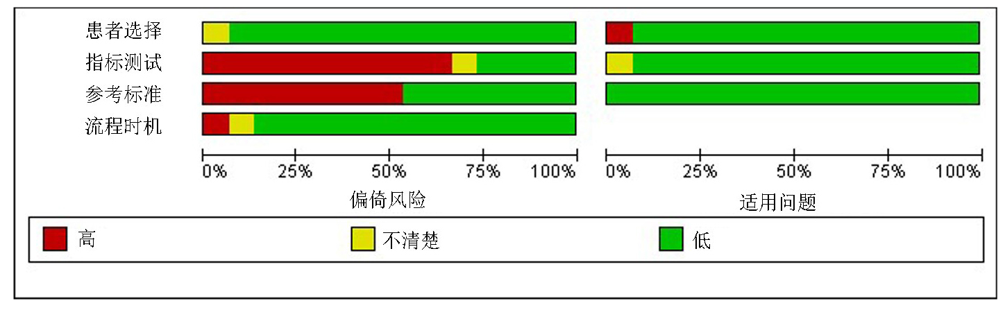
Fig.2 Literature quality assessment matrix
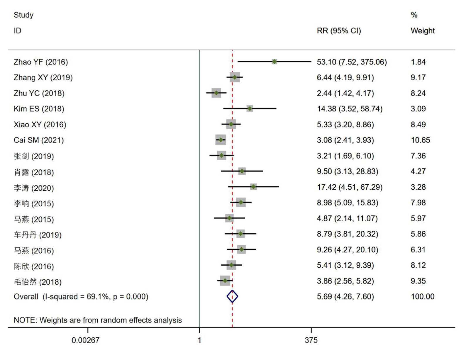
Fig.3 Forest plot and heterogeneity analysis for included 15 papers
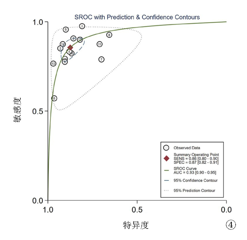
Fig.4 Summary receiver operating characteristic (SROC) curve for superb microvascular imaging
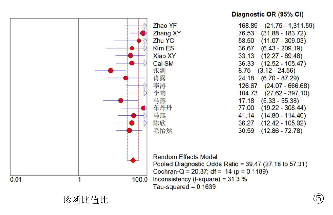
Fig.5 Diagnostic odds ratio (DOR)
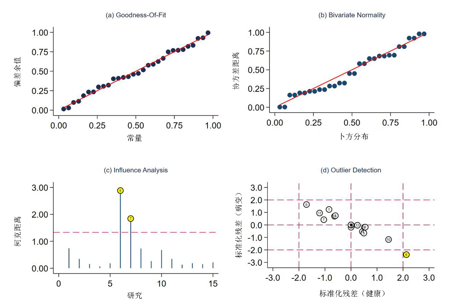
Fig.6 Sensitivity analysis a. Goodness-of-fit test; b. Binary classification test; c. Sensitivity analysis; d. Outliers
| 纳入文献 | 敏感度 95%CI | 特异度 95%CI | 阳性似然比 95%CI | 阴性似然比 95%CI | 诊断比值比 95%CI | SROC曲线下面积 95%CI |
|---|---|---|---|---|---|---|
| 全部文献 | 0.82(0.79~0.84) | 0.87(0.85~0.89) | 6.58(4.58~9.43) | 0.19(0.16~0.24) | 39.47(27.18~57.31) | 0.93(0.90~0.95) |
| 删除6号 | 0.82(0.79~0.85) | 0.87(0.85~0.89) | 6.49(4.44~9.49) | 0.18(0.13~0.26) | 40.37(26.88~60.64) | 0.91(0.88~0.93) |
| 删除7号 | 0.82(0.79~0.84) | 0.88(0.86~0.90) | 7.10(4.90~10.29) | 0.18(0.13~0.25) | 44.11(32.18~60.45) | 0.93(0.91~0.95) |
| 删除6和7 | 0.86(0.83~0.88) | 0.87(0.84~0.89) | 6.68(4.63~9.65) | 0.18(0.15~0.21) | 44.94(32.31~62.50) | 0.93(0.90~0.95) |
Tab.2 Sensitivity analysis results for SMI
| 纳入文献 | 敏感度 95%CI | 特异度 95%CI | 阳性似然比 95%CI | 阴性似然比 95%CI | 诊断比值比 95%CI | SROC曲线下面积 95%CI |
|---|---|---|---|---|---|---|
| 全部文献 | 0.82(0.79~0.84) | 0.87(0.85~0.89) | 6.58(4.58~9.43) | 0.19(0.16~0.24) | 39.47(27.18~57.31) | 0.93(0.90~0.95) |
| 删除6号 | 0.82(0.79~0.85) | 0.87(0.85~0.89) | 6.49(4.44~9.49) | 0.18(0.13~0.26) | 40.37(26.88~60.64) | 0.91(0.88~0.93) |
| 删除7号 | 0.82(0.79~0.84) | 0.88(0.86~0.90) | 7.10(4.90~10.29) | 0.18(0.13~0.25) | 44.11(32.18~60.45) | 0.93(0.91~0.95) |
| 删除6和7 | 0.86(0.83~0.88) | 0.87(0.84~0.89) | 6.68(4.63~9.65) | 0.18(0.15~0.21) | 44.94(32.31~62.50) | 0.93(0.90~0.95) |
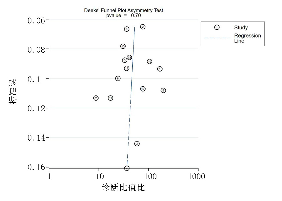
Fig.7 Deeks publication bias test
| [1] |
Gu J, Ternifi R, Larson NB, et al. Hybrid high-definition microvessel imaging/shear wave elastography improves breast lesion characterization[J]. Breast Cancer Res, 2022, 24(1):16.
doi: 10.1186/s13058-022-01511-5 pmid: 35248115 |
| [2] |
Uysal E, Öztürk M, Kilinçer A, et al. Comparison of the effectiveness of shear wave elastography and superb microvascular imaging in the evaluation of breast masses[J]. Ultrasound Q, 2021, 37(2):191-197.
doi: 10.1097/RUQ.0000000000000562 pmid: 34057918 |
| [3] | 王帅. SMI联合BI-RADS-US分类在乳腺癌诊断中的价值研究[J]. 中国临床医学影像杂志, 2020, 31(6):403-405+413. |
| [4] | 罗浩柔, 尹立雪. 超声微血管成像与彩色多普勒血流成像对甲状腺结节诊断价值的Meta分析[J]. 中华医学超声杂志(电子版), 2021, 18(6):554-563. |
| [5] |
Yongfeng Z, Ping Z, Wengang L, et al. Application of a novel microvascular imaging technique in breast lesion evaluation[J]. Ultrasound Med Biol, 2016, 42(9):2097-2105.
doi: 10.1016/j.ultrasmedbio.2016.05.010 pmid: 27321174 |
| [6] |
Zhang XY, Zhang L, Li N, et al. Vascular index measured by smart 3-D superb microvascular imaging can help to differentiate malignant and benign breast lesion[J]. Cancer Manag Res, 2019, 11:5481-5487.
doi: 10.2147/CMAR URL |
| [7] |
Zhu YC, Zhang Y, Deng SH, et al. Evaluation of plasma cell mastitis with superb microvascular imaging[J]. Clin Hemorheol Microcirc, 2019, 72(2):129-138.
doi: 10.3233/CH-180468 URL |
| [8] |
Kim ES, Seo BK, Park EK, et al. Significance of microvascular evaluation of ductal lesions on breast ultrasonography: Influence on diagnostic performance[J]. Clin Imaging, 2018, 51:252-259.
doi: 10.1016/j.clinimag.2018.05.024 URL |
| [9] |
Xiao XY, Chen X, Guan XF, et al. Superb microvascular imaging in diagnosis of breast lesions: A comparative study with contrast-enhanced ultrasonographic microvascular imaging[J]. Br J Radiol, 2016, 89(1066):20160546.
doi: 10.1259/bjr.20160546 URL |
| [10] |
Cai S, Wang H, Zhang X, et al. Superb Microvascular imaging technology can improve the diagnostic efficiency of the BI-RADS system[J]. Front Oncol, 2021, 11:634752.
doi: 10.3389/fonc.2021.634752 URL |
| [11] | 张剑, 陈卉, 徐斌, 等. 超微血管成像、高级动态血流显像、彩色多普勒血流显像对乳腺微小癌的诊断价值及其与病理肿瘤微血管密度的相关性研究[J]. 中华超声影像学杂志, 2019, 28(9):787-793. |
| [12] | 肖露, 褚雯, 王华. 超微血管成像技术对乳腺肿瘤血管形态分特征及其诊断效能的初步分析[J]. 中华超声影像学杂志, 2018, 27(11):973-976. |
| [13] | 李涛, 罗春月, 刘观成, 等. 超声弹性成像联合超微血流成像校正BI-RADS分类的临床价值[J]. 临床超声医学杂志, 2020, 22(8):580-584. |
| [14] | 李响, 康姝, 王学梅, 等. 超微血管成像与彩色多普勒血流成像在乳腺肿瘤诊断中的应用[J]. 中国医学影像技术, 2015, 31(5):663-667. |
| [15] | 马燕, 李刚, 李晶, 等. 超微血管成像技术检测乳腺良恶性肿物血流[J]. 中国医学影像技术, 2015, 31(5):659-662. |
| [16] | 车丹丹, 李玉宏, 于晓溪, 等. 超微血管成像技术对乳腺肿物的诊断价值[J]. 暨南大学学报(自然科学与医学版), 2019, 40(4):352-357. |
| [17] | 马燕, 郭嵩, 李晶, 等. 超微血管成像技术联合超声BI-RADS分级在鉴别乳腺良恶性肿物中应用价值[J]. 中国临床医学影像杂志, 2016, 27(1):10-13. |
| [18] | 陈欣, 肖晓云, 吴欢, 等. 微细血流成像技术在乳腺肿瘤鉴别中的应用[J]. 中国超声医学杂志, 2016, 32(5):407-410. |
| [19] | 毛怡然, 穆洁, 赵静, 等. 超微血管成像与能量多普勒超声对不同大小乳腺实性肿物诊断的对比研究[J]. 中华超声影像学杂志, 2018, 27(4):328-333. |
| [20] | 陈树坤, 祁明. 超微血管成像联合穿刺活检对BI-RADS4类乳腺结节的诊断价值[J]. 锦州医科大学学报, 2022, 43(4):98-102. |
| [21] | 赵利辉, 忻晓洁. 超微血管成像在乳腺及颈部肿瘤中的应用进展[J]. 中国医学影像学杂志, 2021, 29(1):89-92. |
| [22] | 赵荣梅, 姜紫韵, 薛红元, 等. SMI在乳腺导管内乳头状瘤中的诊断价值[J]. 中国超声医学杂志, 2018, 34(11):986-988. |
| [23] | 陈欣, 林伟星, 林健玲, 等. 超微血管成像下乳腺癌的血流特征分析[J]. 中国中西医结合影像学杂志, 2021, 19(3):234-236. |
| [24] | 韩敏, 张宇, 路红, 等. 剪切波弹性成像和超微血管成像辨别乳腺早期恶性病变的价值[J]. 郑州大学学报(医学版), 2022, 57(3):357-361. |
| [25] | 贺烨, 陈卉, 张剑, 等. 超微血管成像技术对BI-RADS 4类乳腺微小肿瘤的诊断价值[J]. 江苏大学学报(医学版), 2020, 30(3):269-271. |
| [26] |
Kayadibi Y, Bulut IN, Aladag Kurt S, et al. The role of superb microvascular imaging and shearwave elastography in the evaluation of intraductal papilloma-like lesions[J]. J Ultrasound Med, 2022, 41(4):995-1008.
doi: 10.1002/jum.v41.4 URL |
| Viewed | ||||||
|
Full text |
|
|||||
|
Abstract |
|
|||||